A new breast lump needs the right level of attention, not an automatic assumption that it is cancer or an automatic decision to remove it. By the end, you will know which symptoms need prompt assessment, how fibroadenoma is confirmed, when observation is reasonable, and when surgery deserves serious consideration.
Key takeaways
- Arrange clinical assessment for every new breast lump.
- A smooth, movable lump cannot confirm fibroadenoma.
- Diagnosis may involve examination, ultrasound, mammography or biopsy.
- Removal depends on symptoms, growth, size and diagnostic certainty.
Which breast lumps need prompt assessment?
A new breast lump needs clinical assessment, even when it feels smooth or movable. Most breast lumps are not cancer, but touch and appearance cannot reliably identify a fibroadenoma, cyst, infection or cancer.
Arrange an urgent breast lump assessment if you notice any of these findings:
- A hard or fixed lump
- Skin dimpling breast lump symptoms, nipple inversion or new nipple retraction
- Spontaneous bloody nipple discharge
- An enlarged lump in the armpit
- Rapid enlargement or a clear change in the breast
- Fever, redness, warmth or severe pain
| Finding | Appointment timing | Why it matters |
|---|---|---|
| Stable, soft or mobile lump without warning signs | Routine appointment soon | It still needs examination and appropriate imaging |
| Hard or fixed lump, rapid growth or armpit lump | Prompt assessment | These features require imaging and often tissue diagnosis |
| Fever, redness, warmth or severe pain | Same-day medical advice | Infection or an abscess may need treatment quickly |
| Bloody discharge, nipple retraction or skin dimpling | Prompt assessment | These changes need investigation rather than observation |
Do not label a lump a fibroadenoma from examination alone. Assessment combines examination, imaging and, when indicated, needle sampling; a benign biopsy is reassuring when it matches the examination and imaging results.
A stable lump without concerning signs can usually enter a routine diagnostic pathway, but do not wait if it grows or develops new changes.
What can cause a breast lump besides cancer?
Most breast lumps are benign, but your fingers cannot reliably identify the cause. A new lump needs clinical assessment with examination, imaging and, when indicated, needle sampling; a reassuring result must agree with both examination and imaging.
Fibroadenoma is a benign solid growth of glandular and fibrous tissue, often mobile, rubbery and found in younger women. A breast cyst contains fluid and may feel smooth or tender, particularly before menstruation. A lipoma in breast tissue is a soft, freely mobile lump made of fat.
| Cause | Typical feel or feature | How it differs |
|---|---|---|
| Fibroadenoma | Firm, rubbery, mobile | Solid benign tissue growth |
| Breast cyst | Smooth, round, sometimes tender | Fluid-filled; ultrasound can show fluid |
| Lipoma in breast | Soft and easily movable | Fatty lump, usually painless |
| Fat necrosis breast lump | Firm, irregular, sometimes after injury or surgery | Damaged fat can mimic cancer on examination or imaging |
| Abscess | Painful, red, warm lump with possible fever | Infection requiring prompt treatment |
| Suspicious solid mass | Hard, fixed, enlarging or irregular | Needs imaging and usually core-needle biopsy |
A papilloma inside a milk duct can cause a small lump or bloody nipple discharge. Rapid enlargement deserves reassessment because a phyllodes tumour can resemble a fibroadenoma. Removing one benign lump also does not prevent a later, different lump from needing investigation.
How doctors diagnose a fibroadenoma
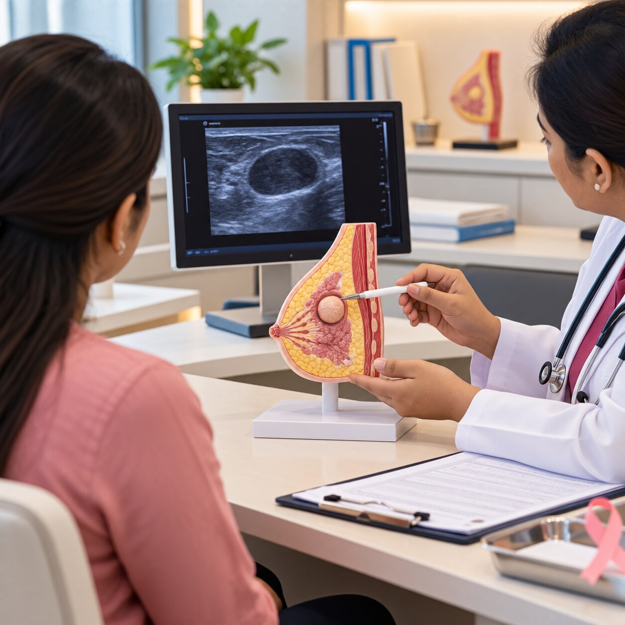
A fibroadenoma cannot be confirmed by touch alone. Doctors combine a clinical breast examination with imaging and, when indicated, needle sampling; the result is reassuring only when the examination, images and pathology agree.
1. Under age 30, breast ultrasound is usually the first imaging test. It can show whether the lump is solid, fluid-filled or has features that need tissue sampling, without exposing young breast tissue to radiation.
2. From age 30 to 39, doctors commonly use breast ultrasound together with diagnostic mammography. The mammogram assesses the wider breast, while targeted ultrasound examines the palpable area in detail.
3. From age 40 onward, diagnostic mammography is usually the starting study, supplemented by targeted ultrasound. Ultrasound remains important because it can characterise a lump that mammography cannot clearly explain.
4. If imaging or examination is suspicious, growing, or discordant, an ultrasound-guided core-needle biopsy is generally preferred to immediate removal. It provides tissue architecture, helping distinguish fibroadenoma from breast cancer and phyllodes tumour.
5. A fine-needle aspiration breast lump procedure removes cells for examination, but it often cannot show the tissue architecture needed to separate fibroadenoma from phyllodes tumour. It is therefore not always sufficient as the only test.
A benign biopsy does not end assessment if the lump feels fixed, enlarges rapidly or looks suspicious on imaging. When examination, imaging and biopsy disagree, doctors reassess the diagnosis and may recommend repeat sampling or excision.
When should a fibroadenoma be removed?
Observation is usually safer than removal when the fibroadenoma is small, stable, confidently identified on imaging, and confirmed by a concordant core-needle biopsy. A planned follow-up examination and imaging schedule lets your clinician detect change without exposing you to a scar, contour change, bleeding or infection.
Do not simply observe fibroadenoma when the lump continues to enlarge, causes pain or pressure, distorts the breast, or creates significant anxiety despite clear counselling. A rapidly growing breast lump deserves reassessment, even after an earlier benign result.
Core-needle biopsy is preferred to immediately removing many lumps because it provides tissue architecture and helps distinguish fibroadenoma from breast cancer and phyllodes tumour. Fine-needle aspiration can sample cells but often cannot make that distinction reliably.
Excision becomes more appropriate when imaging and pathology disagree, the biopsy shows atypical features, or the result does not explain what the clinician or scan found. A core biopsy cannot completely exclude phyllodes tumour in every enlarging or discordant lesion, so removal may be recommended for a definitive diagnosis.
Your informed preference also matters after discussing the benefits, scar and possible breast-shape change.
Removing a benign fibroadenoma does not prevent a later or different lump. Any new change still needs assessment rather than being dismissed as the old lesion returning.
Planning breast lump care in Pune
Before agreeing to fibroadenoma removal Pune, ask the surgeon to review the examination, ultrasound or mammogram, and biopsy report together. A breast surgery consultation should confirm that the findings are benign and match each other, a relationship called radiologic-pathologic concordance.
- What is the exact size, position and imaging appearance of the lump, and has it changed on comparison scans?
- Was a core-needle biopsy performed? Ask why it was chosen over fine-needle aspiration, which often cannot show the tissue architecture needed to distinguish fibroadenoma from phyllodes tumour.
- Does the pathology match the imaging and examination? Discordance, atypical cells or rapid enlargement can justify repeat sampling or excision.
- What is the benefit of removing this lump now compared with surveillance? Ask how often you need clinical review and repeat imaging if it is small, stable and symptom-free.
- Where will the incision sit, and what are the risks of a visible scar, contour change, bleeding, infection, altered sensation or incomplete removal?
- Will the entire specimen go to histopathology, and when will you receive the report?
- What happens if the final result is phyllodes tumour or another unexpected lesion?
A breast lump surgeon Pune patients consults should also explain anaesthesia, expected recovery, activity restrictions and follow-up. Dr Mahesh B Jadhav can help you separate the decision to investigate a lump from the separate decision to operate on it.
Frequently asked questions
Which breast lumps need prompt assessment?
Arrange prompt assessment for any new lump, especially one that is hard, fixed, growing, associated with skin or nipple changes, bloody discharge, an armpit lump or persistent pain.
What can cause a breast lump besides cancer?
Possible causes include fibroadenoma, breast cysts, infection or abscess, fat necrosis, benign breast changes and haematoma after injury.
How do doctors diagnose a fibroadenoma?
A doctor examines the breast and may request an ultrasound, mammogram or core-needle biopsy based on your age, findings and imaging results.
When should a fibroadenoma be removed?
Removal can be considered when the lump grows, causes pain or anxiety, is large, affects breast shape, or remains diagnostically uncertain after testing.